

(Ph. Eur. monograph 1110)
Action and use
Cytokine.
Preparation
Interferon Alfa-2a Injection
DEFINITION
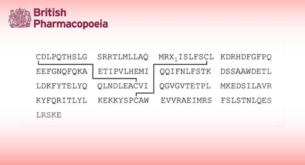
Interferon alfa-2 concentrated solution is a solution of a protein that is produced according to the information coded by the alfa-2 sub-species of interferon alfa gene and that exerts non-specific antiviral activity, at least in homologous cells, through cellular metabolic processes involving synthesis of both ribonucleic acid and protein. Interferon alfa-2 concentrated solution also exerts antiproliferative activity. Different types of alfa-2 interferon, varying in the amino acid residue at position 23, are designated by a letter in lower case.
| Designation | Residue at position 23 (X1) |
| alfa-2a | Lys |
| alfa-2b | Arg |
This monograph applies to interferon alfa-2a and -2b concentrated solutions.
The potency of interferon alfa-2 concentrated solution is not less than 1.4 × 10⁸ IU per milligram of protein. Interferon alfa- 2 concentrated solution contains not less than 2 × 10⁸ IU of interferon alfa-2 per millilitre.
PRODUCTION
Interferon alfa-2 concentrated solution is produced by a method based on recombinant DNA (rDNA) technology using bacteria as host cells. It is produced under conditions designed to minimise microbial contamination of the product.
Interferon alfa-2 concentrated solution complies with the following additional requirements.
Host-cell-derived proteins
The limit is approved by the competent authority.
Host-cell- or vector-derived DNA
The limit is approved by the competent authority.
CHARACTERS
A clear, colourless or slightly yellowish liquid.
IDENTIFICATION
A. It shows the expected biological activity (see Assay).
B. Examine by isoelectric focusing.
Test solution: Dilute the preparation to be examined with water R to a protein concentration of 1 mg/mL.
Reference solution: Prepare a 1 mg/mL solution of interferon alfa-2b CRS in water R.
Isoelectric point calibration solution pI range 3.0 to 10.0 Prepare and use according to the manufacturer’s instructions. Use a suitable apparatus connected with a recirculating temperature controlled water-bath set at 10 °C and gels for isoelectric focusing with a pH gradient from 3.5 to 9.5. Operate the apparatus in accordance with the manufacturer’s instructions. Use as the anode solution phosphoric acid R (98 g/L H3PO4) and as the cathode solution 1 M sodium hydroxide. Samples are applied to the gel by filter papers. Place sample application filters on the gel close to the cathode.
Apply 15 μL of the test solution and 15 μL of the reference solution. Start the isoelectric focusing at 1500 V and 50 mA. Turn off the power after 30 min, remove the application filters and reconnect the power supply for 1 h. Keep the power constant during the focusing process. After focusing, immerse the gel in a suitable volume of a solution containing 115 g/L of trichloroacetic acid R and 34.5 g/L of sulfosalicylic acid R in water R and agitate the container gently for 60 min. Transfer the gel to a mixture of 32 volumes of glacial acetic acid R, 100 volumes of anhydrous ethanol R and 268 volumes of water R, and soak for 5 min. Immerse the gel for 10 min in a staining solution prewarmed to 60 °C in which 1.2 g/L of acid blue 83 R has been added to the previous mixture of glacial acetic acid, ethanol and water. Wash the gel in several containers with the previous mixture of glacial acetic acid, ethanol and water and keep the gel in this mixture until the background is clear (12 h to 24 h). After adequate destaining, soak the gel for 1 h in a 10 per cent V/V solution of glycerol R in the previous mixture of glacial acetic acid, ethanol and water.
The principal bands of the electropherogram obtained with the test solution correspond in position to the principal bands of the electropherogram obtained with the reference solution. Plot the migration distances of the isoelectric point markers versus their isoelectric points and determine the isoelectric points of the principal components of the test solution and the reference solution. They do not differ by more than 0.2 pI units. The test is not valid unless the isoelectric point markers are distributed along the entire length of the gel and the isoelectric points of the principal bands in the electropherogram obtained with the reference solution are between 5.8 and 6.3.
C. Examine the electropherograms obtained under reducing conditions in the test for impurities of molecular masses differing from that of interferon alfa-2. The principal band in the electropherogram obtained with test solution (a) corresponds in position to the principal band in the electropherogram obtained with reference solution (a).
D. Examine by peptide mapping.
Test solution: Dilute the preparation to be examined in water R to a protein concentration of 1.5 mg/mL. Transfer 25 μL to a polypropylene or glass tube of 1.5 mL capacity. Add 1.6 μL of 1 M phosphate buffer solution pH 8.0 R, 2.8 μL of a freshly prepared 1.0 mg/mL solution of trypsin for peptide mapping R in water R and 3.6 μL of water R and mix vigorously. Cap the tube and place it in a water-bath at 37 °C for 18 h, then add 100 μL of a 573 g/L solution of guanidine hydrochloride R and mix well. Add 7 μL of 154.2 g/L solution of dithiothreitol R and mix well. Place the capped tube in boiling water for 1 min. Cool to room temperature.
Reference solution: Prepare at the same time and in the same manner as for the test solution but use a 1.5 mg/mL solution of interferon alfa-2b CRS in water R.
Examine by liquid chromatography (2.2.29).
The chromatographic procedure may be carried out using:
— a stainless steel column 0.10 m long and 4.6 mm in internal diameter packed with octadecylsilyl silica gel for chromatography R (5 μm) with a pore size of 30 nm,
— as mobile phase at a flow rate of 1.0 mL/min:
Mobile phase A: Dilute 1 mL of trifluoroacetic acid R to 1000 mL with water R,
Mobile phase B: To 100 mL of water R add 1 mL of trifluoroacetic acid R and dilute to 1000 mL with acetonitrile for chromatography R,
| Time (min) |
Mobile phase A (per cent V/V) |
Mobile phase B (per cent V/V) |
Comment |
| 0 – 8 | 100 | 0 | isocratic |
| 8 – 68 | 100 → 40 | 0 → 60 | linear gradient |
| 68 – 72 | 40 | 60 | isocratic |
| 72 – 75 | 40 → 100 | 60 → 0 | linear gradient |
| 75 – 80 | 100 | 0 | re-equilibration |
— as detector a spectrophotometer set at 214 nm,
maintaining the temperature of the column at 30 °C.
Equilibrate the column with mobile phase A for at least 15 min.
Inject 100 μL of the test solution and 100 μL of the reference solution.
System suitability: The chromatogram obtained with the reference solution is qualitatively similar to the chromatogram of interferon alfa-2 digest supplied with interferon alfa-2b CRS.
Results: The profile of the chromatogram obtained with the test solution corresponds to that of the chromatogram obtained with the reference solution.
TESTS
Impurities of molecular masses differing from that of interferon alfa-2
Examine by SDS polyacrylamide gel electrophoresis (2.2.31). The test is performed under both reducing and non-reducing conditions, using resolving gels of 14 per cent acrylamide and silver staining as the detection method.
Sample buffer (non-reducing conditions): Mix equal volumes of water R and concentrated SDS-PAGE sample buffer R.
Sample buffer (reducing conditions): Mix equal volumes of water R and concentrated SDS-PAGE sample buffer for reducing conditions R containing 2-mercaptoethanol as the reducing agent.
Test solution (a): Dilute the preparation to be examined in sample buffer to a protein concentration of 0.5 mg/mL.
Test solution (b): Dilute 0.20 mL of test solution (a) to 1 mL with sample buffer.
Reference solution (a): Prepare a 0.625 mg/mL solution of interferon alfa-2b CRS in sample buffer.
Reference solution (b): Dilute 0.20 mL of reference solution (a) to 1 mL with sample buffer.
Reference solution (c): Dilute 0.20 mL of reference solution (b) to 1 mL with sample buffer.
Reference solution (d): Dilute 0.20 mL of reference solution (c) to 1 mL with sample buffer.
Reference solution (e): Dilute 0.20 mL of reference solution (d) to 1 mL with sample buffer.
Reference solution (f): Use a solution of molecular mass standards suitable for calibrating SDS-PAGE gels in the range 15 kDa to 67 kDa.
Place test and reference solutions, contained in covered test-tubes, on a water-bath for 2 min.
Apply 10 μL of reference solution (f) and 50 μL of each of the other solutions to the stacking gel wells. Perform the electrophoresis under the conditions recommended by the manufacturer of the equipment. Detect proteins in the gel by silver staining.
The test is not valid unless: the validation criteria are met (2.2.31); a band is seen in the electropherogram obtained with reference solution (e); and a gradation of intensity of staining is seen in the electropherograms obtained, respectively, with
test solution (a) and test solution (b) and with reference solutions (a) to (e).
The electropherogram obtained with test solution (a) under reducing conditions may show, in addition to the principal band, less intense bands with molecular masses lower than the principal band. No such band is more intense than the principal band in the electropherogram obtained with reference solution (d) (1.0 per cent) and not more than 3 such bands are more intense than the principal band in the electropherogram obtained with reference solution (e) (0.2 per cent).
The electropherogram obtained with test solution (a) under non-reducing conditions may show, in addition to the principal band, less intense bands with molecular masses higher than the principal band. No such band is more intense than the principal band in the electropherogram obtained with reference solution (d) (1.0 per cent) and not more than 3 such bands are more intense than the principal band in the electropherogram obtained with reference solution (e) (0.2 per cent).
Related proteins
Examine by liquid chromatography (2.2.29).
Test solution: Dilute the preparation to be examined with water R to a protein concentration of 1 mg/mL.
0.25 per cent m/m hydrogen peroxide solution Dilute dilute hydrogen peroxide solution R in water R in order to obtain a 0.25 per cent m/m solution.
Reference solution: To a volume of the test solution, add a suitable volume of 0.25 per cent m/m hydrogen peroxide solution to give a final hydrogen peroxide concentration of 0.005 per cent m/m, and allow to stand at room temperature for 1 h, or for the length of time that will generate about 5 per cent oxidised interferon. Add 12.5 mg of L-methionine R per millilitre of solution. Allow to stand at room temperature for 1 h. Store the solutions for not longer than 24 h at a temperature of 2-8 °C.
The chromatographic procedure may be carried out using:
— a stainless steel column 0.25 m long and 4.6 mm in internal diameter packed with octadecylsilyl silica gel for chromatography R (5 μm) with a pore size of 30 nm,
— as mobile phase at a flow rate of 1.0 mL/min:
Mobile phase A To 700 mL of water R add 2 mL of trifluoroacetic acid R and 300 mL of acetonitrile for chromatography R,
Mobile phase B To 200 mL of water R add 2 mL of trifluoroacetic acid R and 800 mL of acetonitrile for chromatography R,
| Time (min) |
Mobile phase A (per cent V/V) |
Mobile phase B (per cent V/V) |
Comment |
| 0 – 1 | 72 | 28 | isocratic |
| 1 – 5 | 72 → 67 | 28 → 33 | linear gradient |
| 5 – 20 | 67 → 63 | 33 → 37 | linear gradient |
| 20 – 30 | 63 → 57 | 37 → 43 | linear gradient |
| 30 – 40 | 57 → 40 | 43 → 60 | linear gradient |
| 40 – 42 | 40 | 60 | isocratic |
| 42 – 50 | 40 → 72 | 60 → 28 | linear gradient |
| 50 – 60 | 72 | 28 | re-equilibration |
— as detector a spectrophotometer set at 210 nm.
Equilibrate the column with the mobile phases in the initial gradient ratio for at least 15 min. Inject 50 μL of each solution.
In the chromatograms obtained, interferon alfa-2 elutes at a retention time of about 20 min. In the chromatogram obtained with the reference solution a peak related to oxidised interferon appears at a relative retention of about 0.9 with reference to the principal peak. The test is not valid unless the resolution between the peaks due to oxidised interferon and interferon is at least 1.0. Consider only the peaks whose relative retention is 0.7 to 1.4 with reference to the principal peak. In the chromatogram obtained with the test solution, the area of any peak, apart from the principal peak, is not greater than 3.0 per cent of the total area of all of the peaks. The sum of the areas of any peaks other than the principal peak is not greater than 5.0 per cent of the total area of all of the peaks.
Bacterial endotoxins (2.6.14)
Less than 100 IU in the volume that contains 1.0 mg of protein.
ASSAY
Protein
Test solution: Dilute the preparation to be examined with water R to obtain a concentration of about 0.5 mg/mL of interferon alfa-2.
Reference solutions: Prepare a stock solution of 0.5 mg/mL of bovine albumin R.
Prepare 8 dilutions of the stock solution containing between 3 μg/mL and 30 μg/mL of bovine albumin R.
Prepare 30-fold and 50-fold dilutions of the test solution. Add 1.25 mL of a mixture prepared the same day by combining 2.0 mL of a 20 g/L solution of copper sulfate pentahydrate R in water R, 2.0 mL of a 40 g/L solution of sodium tartrate R in water R and 96.0 mL of a 40 g/L solution of sodium carbonate R in 0.2 M sodium hydroxide to test-tubes containing 1.5 mL of water R (blank), 1.5 mL of the different dilutions of the test solution or 1.5 mL of the reference solutions. Mix after each addition. After approximately 10 min, add to each test-tube 0.25 mL of a mixture of equal volumes of water R and phosphomolybdotungstic reagent R. Mix after each addition. After approximately 30 min, measure the absorbance (2.2.25) of each solution at 750 nm using the blank as the compensation liquid. Draw a calibration curve from the absorbances of the 8 reference solutions and the corresponding protein contents and read from the curve the content of protein in the test
solution.
Potency
The potency of interferon alfa-2 is estimated by comparing its effect to protect cells against a viral cytopathic effect with the same effect of the appropriate International Standard of human recombinant interferon alfa-2 or of a reference preparation
calibrated in International Units.
The International Unit is the activity contained in a stated amount of the appropriate International Standard. The equivalence in International Units of the International Standard is stated by the World Health Organization.
Carry out the assay by a suitable method, based on the following design.
Use, in standard culture conditions, an established cell line sensitive to the cytopathic effect of a suitable virus (a human diploid fibroblast cell line, free of microbial contamination, responsive to interferon and sensitive to encephalomyocarditis virus, is suitable).
The following cell cultures and virus have been shown to be suitable: MDBK cells (ATCC No. CCL22), or Mouse L cells (NCTC clone 929; ATCC No. CCL 1) as the cell culture and vesicular stomatitis virus VSV, Indiana strain (ATCC No. VR-158) as the infective agent; or A-549 cells (ATCC No. CCL-185) responsive to interferon as the cell culture, and encephalomyocarditis virus (ATCC No. VR-129B) as the infective agent.
Incubate in at least 4 series, cells with 3 or more different concentrations of the preparation to be examined and the reference preparation in a microplate and include in each series appropriate controls of untreated cells. Choose the
concentrations of the preparations such that the lowest concentration produces some protection and the largest concentration produces less than maximal protection against the viral cytopathic effect. Add at a suitable time the cytopathic virus to all wells with the exception of a sufficient number of wells in all series, which are left with uninfected control cells. Determine the cytopathic effect of virus quantitatively with a suitable method. Calculate the potency of the preparation to be examined by the usual statistical methods for a parallel line assay.
The estimated potency is not less than 80 per cent and not more than 125 per cent of the stated potency. The confidence limits of the estimated potency (P = 0.95) are not less than 64 per cent and not more than 156 per cent of the stated potency.
STORAGE
Store in an airtight container, protected from light, at or below -20 °C.
LABELLING
The label states:
— the type of interferon (alfa-2a or alfa-2b),
— the type of production.