


(Ph. Eur. monograph 1912)
DEFINITION
Purified, winterised and deodorised fatty oil obtained from fish of families such as Engraulidae, Carangidae, Clupeidae, Osmeridae, Scombridae and Ammodytidae (type I), or from the genera Thunnus and Sarda within the family Scombridae (type II). The omega-3 acids are defined as the following acids: alpha-linolenic acid (C18:3 n-3), moroctic acid (C18:4 n-3), eicosatetraenoic acid (C20:4 n-3), timnodonic (eicosapentaenoic) acid (C20:5 n-3; EPA), heneicosapentaenoic acid (C21:5 n-3), clupanodonic acid (C22:5 n-3) and cervonic (docosahexaenoic) acid (C22:6 n-3; DHA).
Content
| Type I | Type II | |
| EPA, expressed as triglycerides | minimum 13 per cent | 4 per cent to 12 per cent |
| DHA, expressed as triglycerides | minimum 9 per cent | minimum 20 per cent |
| Total omega-3 acids, expressed as triglycerides |
minimum 28 per cent | minimum 28 per cent |
A suitable antioxidant may be added.
PRODUCTION
The content of dioxins and dioxin-like PCBs (polychlorinated biphenyls) is controlled using methods and limits in accordance with the requirements set in the European Union or other applicable regulations.
CHARACTERS
Appearance
Pale yellow liquid.
Solubility
Practically insoluble in water, very soluble in acetone and in heptane, slightly soluble in anhydrous ethanol.
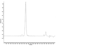
1. oligomers 2. triglycerides
Figure 1912.-1. – Chromatogram for the test for oligomers in fish oil rich in omega-3 acids
IDENTIFICATION
A. Examine the chromatograms obtained in the assay for EPA and DHA.
Results: The peaks due to eicosapentaenoic acid methyl ester and docosahexaenoic acid methyl ester in the chromatogram obtained with test solution (b) are similar in retention time to the corresponding peaks in the chromatogram obtained with reference solutions (a1) and (a2).
B. It complies with the limits of the assay for EPA (type I or II).
TESTS
Appearance
The substance to be examined is not more intensely coloured than a reference solution prepared as follows: to 3.0 mL of red primary solution add 25.0 mL of yellow primary solution and dilute to 50.0 mL with a 10 g/L solution of hydrochloric acid R (2.2.2, Method II).
Absorbance (2.2.25)
Maximum 0.70 (type I) or maximum 0.50 (type II), at 233 nm.
Dilute 0.300 g of the substance to be examined to 50.0 mL with trimethylpentane R. Dilute 2.0 mL of the solution to 50.0 mL with trimethylpentane R.
Acid value (2.5.1)
Maximum 0.5, determined on 20.0 g in 50 mL of a mixture of equal volumes of ethanol (96 per cent) R and ether R.
Anisidine value (2.5.36)
Maximum 30.0 (type I) or maximum 15.0 (type II).
Peroxide value (2.5.5, Method A)
Maximum 10.0 (type I) or maximum 5.0 (type II).
Unsaponifiable matter (2.5.7)
Maximum 1.5 per cent, determined on 5.0 g.
Stearin
Immerse at least 10 mL of the substance to be examined previously at room temperature, for 3 h in a bath of iced water or a thermostatically controlled bath at 0 ± 0.5 °C. The sample remains clear.
Oligomers
Size-exclusion chromatography (2.2.30).
Test solution: Dilute 50.0 mg of the substance to be examined to 10.0 mL with tetrahydrofuran R.
Reference solution: In a 100 mL volumetric flask, dissolve 50 mg of monodocosahexaenoin R, 30 mg of didocosahexaenoin R and 20 mg of tridocosahexaenoin R in tetrahydrofuran R and dilute to 100.0 mL with the same solvent.
Column 3 columns to be connected in series:
— size: l = 0.3 m, Ø = 7.8 mm;
— stationary phase: styrene-divinylbenzene copolymer R (5 μm) with the following pore sizes:
— column 1: 50 nm;
— column 2: 10 nm;
— column 3: 5 nm;
— connection sequence: injector – column 1 – column 2 – column 3 – detector.
Mobile phase: tetrahydrofuran R.
Flow rate: 0.8 mL/min.
Detection: Differential refractometer.
Injection: 40 μL.
System suitability: Reference solution:
— elution order: tridocosahexaenoin, didocosahexaenoin, monodocosahexaenoin;
— resolution: minimum 2.0 between the peaks due to didocosahexaenoin and monodocosahexaenoin and minimum 1.0 between the peaks due to tridocosahexaenoin and didocosahexaenoin.
Identify the peaks from the chromatogram (Figure 1912.-1). Calculate the percentage content of oligomers using the following expression:
A/B x 100
A = sum of the areas of all the peaks in the chromatogram;
B = area of the peak with a retention time less than the retention time of the triglyceride peak.
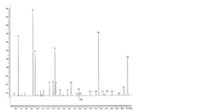
1. C14:0; 2. C16:0; 3. C16:1 n-7; 4. C16:4 n-1; 5. C18:0; 6. C18:1 n-9; 7. C18:1 n-7; 8. C18:2 n-6; 9. C18:3 n-3; 10. C18:4 n-3; 11. C20:0; 12. C20:1 n-9; 12a. C20:1 n-11; 13. C20:1 n-7; 14. C20:4 n-6; 15. C20:4 n-3; 16. C20:5 n-3; 17. C22:1 n-11; 18. C22:1 n-9; 19. C21:5 n-3; 20. C22:5 n-6; 21. C22:5 n-3; 22. C22:6 n-3
Figure 1912.-2. – Chromatogram for the assay of total omega-3 acids in fish oil rich in omega-3 acids
Limit:
— oligomers: maximum 1.5 per cent.
ASSAY
EPA and DHA (2.4.29)
For identification of the peaks, see Figure 1912.-2.
Total omega-3 acids (2.4.29)
See Figure 1912.-2.
STORAGE
Under an inert gas, in a well-filled, airtight container, protected from light.
LABELLING
The label states:
— the concentration of EPA, DHA and total omega-3 acids, expressed as triglycerides;
— the type of fish oil rich in omega-3 acids (type I or II).