

(Ph. Eur. monograph 1316)
The label states (1) the type of erythropoeitin using the appropriate International Nonproprietary Name (Epoetin Alfa, Epoetin Beta, etc) and (2) the approved code in lower case letters indicative of the method of production.
Mr approx. 30 600
Action and use
Erythropoietin analogue.
Preparation
Erythropoietin Injection
DEFINITION
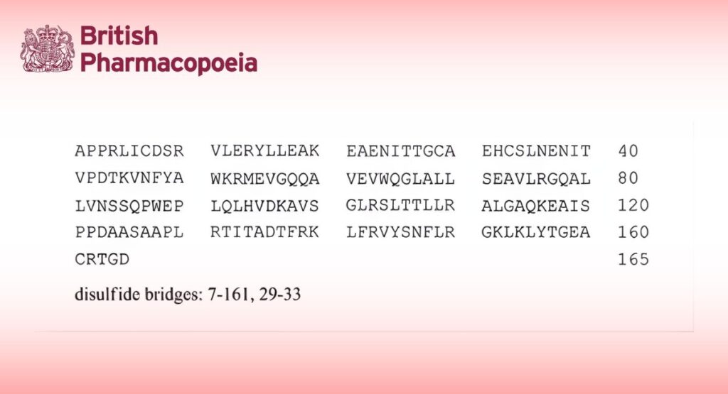
Solution containing a family of closely-related glycoproteins which are indistinguishable from the naturally occurring human erythropoietin (urinary erythropoietin) in terms of amino acid sequence (165 amino acids), at a concentration of 0.5-10 mg/mL. It may also contain buffer salts and other excipients. It has a potency of not less than 100 000 IU per milligram of active substance determined using the conditions described under Assay and in the test for protein.
PRODUCTION
Erythropoietin is produced in rodent cells in vitro by a method based on recombinant DNA (rDNA) technology. During the course of product development, it must be demonstrated that the manufacturing process consistently produces a product with the expected glycosylation pattern using suitably qualified assay(s).
Prior to batch release, the following tests are carried out on each batch of the erythropoietin concentrated solution, unless exemption has been granted by the competent authority.
Host cell-derived proteins
The limit is approved by the competent authority.
Host cell- and vector-derived DNA
The limit is approved by the competent authority.
Glycan analysis
Use a suitable method developed according to general chapter 2.2.59. Glycan analysis of glycoproteins, section 2-3:
— after desalting, release the N–glycans using 1 of the agents described in Table 2.2.59.-1, for example peptide N-glycosidase F (PNGase F);
— if needed, label the released N–glycans with 1 of the fluorescent labelling agents described in Table 2.2.59.-2;
— analyse the labelled or unlabelled N-glycans using a suitable technique.
The following procedure is given as an example.
Solution A: Dissolve 0.77 g of ammonium acetate R in water R and dilute to 1000.0 mL with the same solvent.
Test solution: Desalt a volume of the preparation to be examined by a suitable method (for example, using a suitable centrifugal filter) with solution A. Dilute with solution A to obtain a concentration of approximately 1 mg/mL. Add 500 U of peptide N-glycosidase F R per 100 μL of the obtained solution and incubate at 37 °C for at least 18 h. Evaporate the solvent completely by vacuum centrifugation at room temperature. Reconstitute the dried material by adding 80 μL water R per 100 μL of deglycosylated erythropoietin.
Reference solution (a): Dissolve the contents of a vial of erythropoietin for physicochemical tests CRS in water R. Desalt a volume of this preparation and carry out N-glycan release at the same time and in the same manner as for the test solution.
Reference solution (b): Use a suitable erythropoietin in-house reference preparation shown to be representative of batches tested clinically and batches used to demonstrate consistency of production. Desalt a volume of this preparation and carry out the N-glycan release at the same time and in the same manner as for the test solution.
Analyse the native N-glycans by liquid chromatography (2.2.29).
A precolumn containing strongly basic anion-exchange resin for chromatography R2 (8.5 μm), may be used.
Column:
— size: l = 0.25 m, Ø = 4.0 mm;
— stationary phase: strongly basic anion-exchange resin for chromatography R2 (8.5 μm);
— temperature: 30 °C.
Mobile phase:
— mobile phase A: water for chromatography R;
— mobile phase B: 40 g/L solution of sodium hydroxide R;
— mobile phase C: 82 g/L solution of anhydrous sodium acetate R;
| Time
(min) |
Mobile phase A
(per cent V/V) |
Mobile phase B
(per cent V/V) |
Mobile phase C
(per cent V/V) |
| 0 – 60 | 85 → 67 | 10 | 5 → 23 |
| 60 – 61 | 67 → 0 | 10 → 25 | 23 → 75 |
| 61 – 65 | 0 | 25 | 75 |
| 65 – 66 | 0 → 85 | 25 → 10 | 75 → 5 |
| 66 – 80 | 85 | 10 | 5 |
Flow rate: 1.0 mL/min
Detection: Electrochemical detector (pulsed amperometry).
Autosampler: Set at 10 °C.
Injection: 50 μL.
Identification of peaks: Use the chromatogram supplied with erythropoietin for physicochemical tests CRS to identify the 4 peak clusters corresponding to mono- (S1), bi- (S2), tri- (S3) and tetra-sialylated (S4) N-glycans.
System suitability:
— the chromatogram obtained with reference solution (a) is qualitatively similar to the chromatogram supplied with erythropoietin for physicochemical tests CRS.
Results:
— the profile of the chromatogram obtained with the test solution corresponds to that of the chromatogram obtained with reference solution (b);
— the retention times of the peaks in the chromatogram obtained with the test solution correspond to those in the chromatogram obtained with reference solution (b);
— no additional peaks are observed in the chromatogram obtained with the test solution in comparison with the chromatogram obtained with reference solution (b).
Calculate the percentage contents of mono-, bi-, tri- and tetra-sialylated N-glycans taking into account the total relative peak area for each peak cluster with reference to the sum of the areas of all peak clusters.
Limits:
— percentage of mono-sialylated N-glycans: as authorised by the competent authority;
— percentage of bi-sialylated N-glycans: as authorised by the competent authority;
— percentage of tri-sialylated N-glycans: as authorised by the competent authority;
— percentage of tetra-sialylated N-glycans: as authorised by the competent authority.
CHARACTERS
Appearance
Clear or slightly turbid, colourless solution.
IDENTIFICATION
A. It gives the appropriate response when examined using the conditions described under Assay.
B. Capillary zone electrophoresis (2.2.47).
Test solution: Dilute the preparation to be examined with water R to obtain a concentration of 1 mg/mL. Desalt 0.25 mL of the solution by passage through a micro-concentrator cartridge provided with a membrane with a molecular mass cut-off of not more than 10 000 Da. Add 0.2 mL of water R to the sample and desalt again.
Repeat the desalting procedure once more. Dilute the sample with water R, determine its protein concentration as described under Tests and adjust to a concentration of approximately 1 mg/mL with water R.
Reference solution: Dissolve the contents of a vial of erythropoietin for physicochemical tests CRS in 0.10 mL of water R.
Proceed with desalting as described for the test solution.
Capillary:
— material: uncoated fused silica;
— size: effective length = about 100 cm, Ø = 50 μm.
Temperature 35 °C.
CZE buffer concentrate (0.1 M sodium chloride, 0.1 M tricine, 0.1 M sodium acetate): Dissolve 0.584 g of sodium chloride R, 1.792 g of tricine R and 0.820 g of anhydrous sodium acetate R in water R and dilute to 100.0 mL with the same solvent.
1 M putrescine solution Dissolve 0.882 g of putrescine R in 10 mL of water R. Distribute in 0.5 mL aliquots.
CZE buffer (0.01 M tricine, 0.01 M sodium chloride, 0.01 M sodium acetate, 7 M urea, 2.5 mM putrescine): Dissolve 21.0 g of urea R in 25 mL of water R by warming in a water-bath at 30 °C. Add 5.0 mL of CZE buffer concentrate and 125 μL of 1 M putrescine solution. Dilute to 50.0 mL with water R. Using dilute acetic acid R, adjust to pH 5.55 at 30 °C and filter through a membrane filter (nominal pore size 0.45 μm).
Detection: Spectrophotometer at 214 nm.
Set the autosampler to store the samples at 4 °C during analysis.
Preconditioning of the capillary: Rinse the capillary for 60 min with 0.1 M sodium hydroxide filtered through a membrane filter (nominal pore size 0.45 μm) and for 60 min with CZE buffer. Apply voltage for 12 h (20 kV).
Between-run rinsing: Rinse the capillary for 10 min with water R, for 5 min with 0.1 M sodium hydroxide filtered through a membrane filter (nominal pore size 0.45 μm) and for 10 min with CZE buffer.
Injection: Under pressure or vacuum.
Migration: Apply a field strength of 143 V/cm (15.4 kV for capillaries of 107 cm total length) for 80 min, using CZE buffer as the electrolyte in both buffer reservoirs.
System suitability: In the electropherogram obtained with the reference solution, a pattern of well separated peaks corresponding to the peaks in the electropherogram of erythropoietin supplied with erythropoietin for physicochemical tests CRS is seen, and the largest peak is at least 50 times greater than the baseline noise. If necessary, adjust the sample load to give peaks of sufficient height. Identify the peaks due to isoforms 1 to 8. Isoform 1 may not be visible. The peak due to isoform 8 is detected and the resolution between the peaks due to isoforms 5 and 6 is not less than 1. Repeat the separation at least 3 times. The baseline is stable, showing little drift, and the distribution of peaks is qualitatively and quantitatively similar to the distribution of peaks in the electropherogram of erythropoietin supplied with erythropoietin for physicochemical tests CRS. The relative standard deviation of the migration time of the peak due to isoform 2 is less than 2 per cent.
Limits: Identify the peaks due to isoforms 1 to 8 in the electropherogram obtained with the test solution by comparison with the electropherogram obtained with the reference solution. Calculate the percentage content of each isoform from the corresponding peak area. The percentages are within the following ranges:
| Isoform | Content (per cent) |
| 1 | 0 – 15 |
| 2 | 0 – 15 |
| 3 | 1 – 20 |
| 4 | 10 – 35 |
| 5 | 15 – 40 |
| 6 | 10 – 35 |
| 7 | 5 – 25 |
| 8 | 0 – 15 |
C. Polyacrylamide gel electrophoresis and immunoblotting.
(a) Polyacrylamide gel electrophoresis (2.2.31).
Gel dimensions 0.75 mm thick, about 16 cm square.
Resolving gel 12 per cent acrylamide.
Sample buffer concentrated SDS-PAGE sample buffer R.
Test solution (a): Dilute the preparation to be examined in water R to obtain a concentration of 1.0 mg/mL. To 1 volume of this solution add 1 volume of sample buffer.
Test solution (b): Dilute the preparation to be examined in water R to obtain a concentration of 0.1 mg/mL. To 1 volume of this solution add 1 volume of sample buffer.
Reference solution (a): Dissolve the contents of a vial of erythropoietin for physicochemical tests CRS in 0.10 mL of water R. To 1 volume of this solution add 1 volume of sample buffer.
Reference solution (b): Dissolve the contents of a vial of erythropoietin for physicochemical tests CRS in water R and dilute with the same solvent to obtain a concentration of 0.1 mg/mL. To 1 volume of this solution add 1 volume of sample buffer.
Reference solution (c): A solution of molecular mass markers suitable for calibrating SDS-polyacrylamide gels in the range of 10-70 kDa.
Reference solution (d) A solution of pre-stained molecular mass markers suitable for calibrating SDS-polyacrylamide gels in the range of 10-70 kDa and suitable for the electrotransfer to an appropriate membrane.
Sample treatment Boil for 2 min.
Application: 20 μL, in the following order: reference solution (c), reference solution (a), test solution (a), empty well, reference solution (b), test solution (b), reference solution (d).
At the end of the separation, remove the gel-cassette from the apparatus and cut the gel into 2 parts: the first part containing reference solution (c), reference solution (a) and test solution (a); the second part containing reference solution (b), test solution (b) and reference solution (d).
Detection: By Coomassie staining on the first part of the gel.
System suitability: Reference solution (c):
— the validation criteria are met.
Results: The electropherogram obtained with test solution (a) shows a single diffuse band corresponding in position and intensity to the single band seen in the electropherogram obtained with reference solution (a).
(b) Immunoblotting.
Transfer the second part of the gel onto a membrane suitable for the immobilisation of proteins, using commercially available electrotransfer equipment and following the manufacturer’s instructions. After electrotransfer, incubate the membrane in a neutral isotonic buffer containing a suitable blocking agent (for example, 50 g/L of dried milk or 10 per cent V/V foetal calf serum), for 1-2 h, followed by incubation for 1-14 h in the same blocking solution with a suitable dilution of either a polyclonal or monoclonal anti-erythropoietin antibody. Detect erythropoietin-bound antibody using a suitable enzyme- or radiolabelled antibody (for example, an alkaline phosphatase-conjugated second antibody). The precise details of blocking agents, concentrations and incubation times should be optimised using the principles set out in general chapter (2.7.1) Immunochemical methods.
System suitability: In the electropherogram obtained with reference solution (d), the molecular mass markers are resolved on the membrane into discrete bands, with a linear relationship between distance migrated and logarithm10 of the molecular mass.
Results: The electropherogram obtained with test solution (b) shows a single broad band corresponding in position and intensity to the single band seen in the electropherogram obtained with reference solution (b).
D. Peptide mapping (2.2.55).
SELECTIVE CLEAVAGE OF THE PEPTIDE BONDS
Test solution: Dilute the preparation to be examined in tris acetate buffer solution pH 8.5 R to a concentration of 1.0 mg/mL. Equilibrate the solution in tris acetate buffer solution pH 8.5 R using a suitable procedure (dialysis against tris acetate buffer solution pH 8.5 R, or membrane filtration using the procedure described under Identification B, but reconstituting the desalted sample with tris acetate buffer solution pH 8.5 R, are suitable).
Transfer the dialysed solution to a polypropylene centrifuge tube. Freshly prepare a solution of trypsin for peptide mapping R at a concentration of 1 mg/mL in water R, and add 2 μL to 0.10 mL of the dialysed solution. Cap the tube and place in a water-bath at 37 °C for 18 h.
Remove the sample from the water-bath and stop the reaction immediately by freezing.
Reference solution: Dissolve the contents of a vial of erythropoietin for physicochemical tests CRS in 0.10 mL of water R.
Prepare as for the test solution, ensuring that all procedures are carried out simultaneously, and under identical conditions.
CHROMATOGRAPHIC SEPARATION
Liquid chromatography (2.2.29).
Column:
— size: l = 0.25 m, Ø = 4.6 mm;
— stationary phase: end-capped butylsilyl silica gel for chromatography R (5-10 μm).
Mobile phase:
— mobile phase A: 0.06 per cent V/V solution of trifluoroacetic acid R;
— mobile phase B: to 100 mL of water for chromatography R add 0.6 mL of trifluoroacetic acid R and dilute to 1000 mL with acetonitrile R1;
| Time
(min) |
Flow rate
(mL/min) |
Mobile phase A
(per cent V/V) |
Mobile phase B
(per cent V/V) |
| 0 – 60 | 0.75 | 100 | 0 |
| 60 – 61 | 0.75 | 100 → 39 | 0 → 61 |
| 61 – 65 | 1.25 | 39 → 17 | 61 → 83 |
| 65 – 66 | 1.25 | 17 → 100 | 83 → 100 |
| 66 – 80 | 1.25 | 100 | 100 |
Detection: Spectrophotometer at 214 nm.
Equilibration: At initial conditions for at least 15 min. Carry out a blank run using the above-mentioned gradient.
Injection: 50 μL.
System suitability: The chromatogram obtained with the reference solution is qualitatively similar to the chromatogram of erythropoietin digest supplied with erythropoietin for physicochemical tests CRS.
Results: The profile of the chromatogram obtained with the test solution corresponds to that of the chromatogram obtained with the reference solution.
TESTS
Protein (2.5.33, Method I)
80 per cent to 120 per cent of the stated concentration.
Test solution: Dilute the preparation to be examined in a 4 g/L solution of ammonium hydrogen carbonate R.
Record the absorbance spectrum between 250 nm and 400 nm. Measure the value at the absorbance maximum (276- 280 nm), after correction for any light scattering, measured up to 400 nm. Calculate the concentration of erythropoietin taking the specific absorbance to be 7.43.
Dimers and related substances with molecular masses greater than that of erythropoietin
Size-exclusion chromatography (2.2.30): use the normalisation procedure.
Test solution: Dilute the preparation to be examined in the mobile phase to obtain a concentration of 0.2 mg/mL.
Resolution solution: Dissolve the contents of a vial of erythropoietin for SEC system suitability CRS in the mobile phase to obtain a concentration of 0.2 mg/mL.
Reference solution: Dilute 20 μL of the resolution solution to 1.0 mL with the mobile phase.
Column:
— size: l = 0.3 m, Ø = 7.8 mm;
— stationary phase: hydrophilic silica gel for chromatography R (5 μm), of a grade suitable for fractionation of globular proteins in the relative molecular mass range of 20 000 to 200 000.
Mobile phase: Dissolve 1.15 g of anhydrous disodium hydrogen phosphate R, 0.2 g of potassium dihydrogen phosphate R and 23.4 g of sodium chloride R in 900 mL of water for chromatography R, adjust to pH 7.4 if necessary, and dilute to 1000 mL with water for chromatography R.
Flow rate: 0.5 mL/min.
Detection: Spectrophotometer at 214 nm.
Injection: 100 μL.
Run time: Twice the retention time of erythropoietin monomer.
Relative retention: With reference to erythropoietin monomer: erythropoietin dimer = about 0.9.
System suitability:
— the area of the principal peak in the chromatogram obtained with the reference solution is 1.5 per cent to 2.5 per cent of the area of the principal peak in the chromatogram obtained with the resolution solution;
— resolution: minimum 1.5 between the peaks due to erythropoietin dimer and erythropoietin monomer in the chromatogram obtained with the resolution solution.
Limit:
— sum of the peaks eluted before the principal peak: maximum 2.0 per cent; disregard any peak with a retention time greater than that of the principal peak.
Sialic acids
Minimum 10 mol of sialic acids (calculated as N-acetylneuraminic acid) per mole of erythropoietin.
Test solution (a): Dilute the preparation to be examined in the mobile phase used in the test for dimers and related substances with molecular masses greater than that of erythropoietin to obtain a concentration of 0.3 mg/mL.
Test solution (b): To 0.5 mL of test solution (a) add 0.5 mL of the mobile phase used in the test for dimers and related substances with molecular masses greater than that of erythropoietin.
Reference solution (a): Dissolve a suitable amount of N-acetylneuraminic acid R in water R to obtain a concentration of 0.1 mg/mL.
Reference solution (b): To 0.8 mL of reference solution (a) add 0.2 mL of water R.
Reference solution (c): To 0.6 mL of reference solution (a) add 0.4 mL of water R.
Reference solution (d): To 0.4 mL of reference solution (a) add 0.6 mL of water R.
Reference solution (e): To 0.2 mL of reference solution (a) add 0.8 mL of water R.
Reference solution (f): Use water R.
Carry out the test in triplicate. Transfer 100 μL of each of the test and reference solutions to 10 mL glass test tubes. To each tube add 1.0 mL of resorcinol reagent R. Stopper the tubes and incubate at 100 °C for 30 min. Cool on ice. To each tube, add 2.0 mL of a mixture of 12 volumes of butanol R and 48 volumes of butyl acetate R. Mix vigorously, and allow the 2 phases to separate. Ensuring that the upper phase is completely clear, remove the upper phase, taking care to exclude completely any of the lower phase.
Measure the absorbance (2.2.25) of all samples at 580 nm. Using the calibration curve generated by the reference solutions, determine the content of sialic acids in test solutions (a) and (b) and calculate the mean. Calculate the number of moles of sialic acids per mole of erythropoietin assuming that the relative molecular mass of erythropoietin is 30 600 and that the relative molecular mass of N-acetylneuraminic acid is 309.
System suitability:
— the individual replicates agree to within ± 10 per cent of each other;
— the value obtained from reference solution (a) is between 1.5 and 3.3 times that obtained with test solution (a).
Bacterial endotoxins (2.6.14)
Less than 20 IU in the volume that contains 100 000 IU of erythropoietin.
ASSAY
The activity of the preparation is compared with that of erythropoietin BRP and expressed in International Units (IU).
The estimated potency is not less than 80 per cent and not more than 125 per cent of the stated potency. The confidence limits of the estimated potency (P = 0.95) are not less than 64 per cent and not more than 156 per cent of the stated potency.
Carry out the determination of potency by Method A or B.
A. In polycythaemic mice
The activity of the preparation is estimated by examining, under given conditions, its effect in stimulating the incorporation of Fe into circulating red blood cells of mice made polycythaemic by exposure to reduced atmospheric pressure.
The following schedule, using treatment in a hypobaric chamber, has been found to be suitable.
Induce polycythaemia in female mice of the same strain, weighing 16-18 g. Place the mice in a hypoxic chamber and reduce the pressure to 0.6 atmospheres. After 3 days at 0.6 atmospheres, further reduce the pressure to 0.4-0.5 atmospheres and maintain the animals at this pressure for a further 11 days (the partial vacuum is interrupted daily for a maximum of 1 h at about 11:00 a.m., in order to clean the cages and feed the animals). At the end of the specified period, return the mice to normal atmospheric conditions.
Randomly distribute the mice into cages, each containing 6 animals, and mark them.
Test solution (a): Dilute the substance to be examined in phosphate-albumin buffered saline pH 7.2 R1 to obtain a concentration of 0.2 IU/mL.
Test solution (b): Mix equal volumes of test solution (a) and phosphate-albumin buffered saline pH 7.2 R1.
Test solution (c): Mix equal volumes of test solution (b) and phosphate-albumin buffered saline pH 7.2 R1.
Reference solution (a): Dissolve erythropoietin BRP in phosphate-albumin buffered saline pH 7.2 R1 to obtain a concentration of 0.2 IU/mL.
Reference solution (b): Mix equal volumes of reference solution (a) and phosphate-albumin buffered saline pH 7.2 R1.
Reference solution (c) Mix equal volumes of reference solution (b) and phosphate-albumin buffered saline pH 7.2 R1.
Radiolabelled ferric [ Fe] chloride solution, concentrated Use a commercially available solution of [ Fe]ferric chloride (approximate specific activity: 100-1000 MBq/mg of Fe).
Radiolabelled [ Fe] ferric chloride solution Dilute the concentrated radiolabelled [ Fe]ferric chloride solution in Sodium citrate buffer solution pH 7.8 (0.034 M sodium citrate, 0.101 M sodium chloride) R to obtain a solution with an activity of 3.7 × 10 Bq/mL.
The concentrations of the test solutions and reference solutions may need to be modified, based on the response range of the animals used.
3 days after returning the animals to atmospheric pressure, inject each animal subcutaneously with 0.2 mL of one of the solutions. The 6 animals in each cage must each receive one of the 6 different treatments (3 test solutions and 3 reference solutions), and the order of injection must be separately randomised for each cage. A minimum of 8 cages is recommended. 2 days after injection of the test or reference solution, inject each animal intraperitoneally with 0.2 mL of radiolabelled [ Fe]ferric chloride solution. The order of the injections must be the same as that of the erythropoietin injections, and the time interval between administration of the erythropoietin and the radiolabelled ferric chloride solution must be the same for each animal. After a further 48 h, anaesthetise each animal by injection of a suitable anaesthetic, record body weights and withdraw blood samples (0.65 mL) into haematocrit capillaries from the bifurcation of the aorta. After determining the packed cell volume for each sample, measure the radioactivity.
Calculate the response (percentage of iron-59 in total circulating blood) for each mouse using the expression:
(As x M x 7.5)/ (At x Vs)
As = radioactivity in the sample;
At = total radioactivity injected;
7.5 = total blood volume as per cent body weight;
M = body weight, in grams;
Vs = sample volume.
Calculate the potency by the usual statistical methods for a parallel line assay. Eliminate from the calculation any animal where the packed cell volume is less than 54 per cent, or where the body weight is more than 24 g.
B. In normocythaemic mice
The assay is based on the measurement of stimulation of reticulocyte production in normocythaemic mice.
The assay may be carried out using the following procedure:
Test solution (a): Dilute the preparation to be examined in phosphate-albumin buffered saline pH 7.2 R1 to obtain a concentration of 80 IU/mL.
Test solution (b): Mix equal volumes of test solution (a) and phosphate-albumin buffered saline pH 7.2 R1.
Test solution (c): Mix equal volumes of test solution (b) and phosphate-albumin buffered saline pH 7.2 R1.
Reference solution (a): Dissolve erythropoietin BRP in phosphate-albumin buffered saline pH 7.2 R1 to obtain a concentration of 80 IU/mL.
Reference solution (b) Mix equal volumes of reference solution (a) and phosphate-albumin buffered saline pH 7.2 R1.
Reference solution (c) Mix equal volumes of reference solution (b) and phosphate-albumin buffered saline pH 7.2 R1.
The exact concentrations of the test solutions and reference solutions may need to be modified, based on the response range of the animals used.
At the beginning of the assay procedure, randomly distribute mice of a suitable age and strain (8-week old B6D2F1 mice are suitable) into 6 cages. A minimum of 8 mice per cage is recommended. Inject each animal subcutaneously with 0.5 mL of the appropriate treatment (one solution per cage) and put the animal in a new cage. Combine the mice in such a way that each cage housing the treated mice contains one mouse out of the 6 different treatments (3 test solutions and 3 reference solutions, 6 mice per cage). 4 days after the injections, collect blood samples from the animals and determine the number of reticulocytes using a suitable procedure.
The following method may be employed:
The volume of blood, dilution procedure and fluorescent reagent may need to be modified to ensure maximum development and stability of fluorescence.
Colorant solution, concentrated Use a solution of thiazole orange suitable for the determination of reticulocytes. Prepare at a concentration twice that necessary for the analysis.
Proceed with the following dilution steps. Dilute whole blood 500-fold in the buffer used to prepare the colorant solution.
Dilute this solution 2-fold in the concentrated colorant solution. After staining for 3-10 min, determine the reticulocyte count microfluorometrically in a flow cytometer. The percentage of reticulocytes is determined using a biparametric histogram: number of cells/red fluorescence (620 nm).
Calculate the potency by the usual statistical methods for a parallel line assay.
STORAGE
In an airtight container at a temperature below -20 °C. Avoid repeated freezing and thawing.
LABELLING
The label states:
— the erythropoietin content in milligrams per millilitre;
— the activity in International Units per millilitre;
— the name and the concentration of any other excipients.